Breakthrough in Cellular Imaging: SPIFFI Technology Enables Single-Frame Super-Resolution Microscopy for Live Cells

By Science and Technology Desk
Published in partnership with advanced research briefings
Main Facts
In a monumental leap forward for cellular biology and optical engineering, a team of researchers has unveiled a novel fluorescence microscopy technique capable of generating high-contrast, super-resolution images from a single exposure. Dubbed SPIFFI—which stands for spatial polarization-induced fluorescence fluctuation imaging—the technology was developed by scientists Wei Guo, Lely Feletti, and Aleksandra Radenovic at the Laboratory of Nanoscale Biology (LBEN) within the École Polytechnique Fédérale de Lausanne (EPFL) School of Engineering.
Published in the esteemed scientific journal Nature, the breakthrough addresses one of the most stubborn bottlenecks in optical microscopy: the inability to capture high-resolution imagery of dynamic, living cellular processes without motion blur or phototoxicity.
Traditional super-resolution microscopy methods typically rely on capturing hundreds or even thousands of sequential frames to synthesize a single, high-resolution image. While revolutionary for static samples, this temporal accumulation strategy fails catastrophically when applied to living cells, which are perpetually in motion.
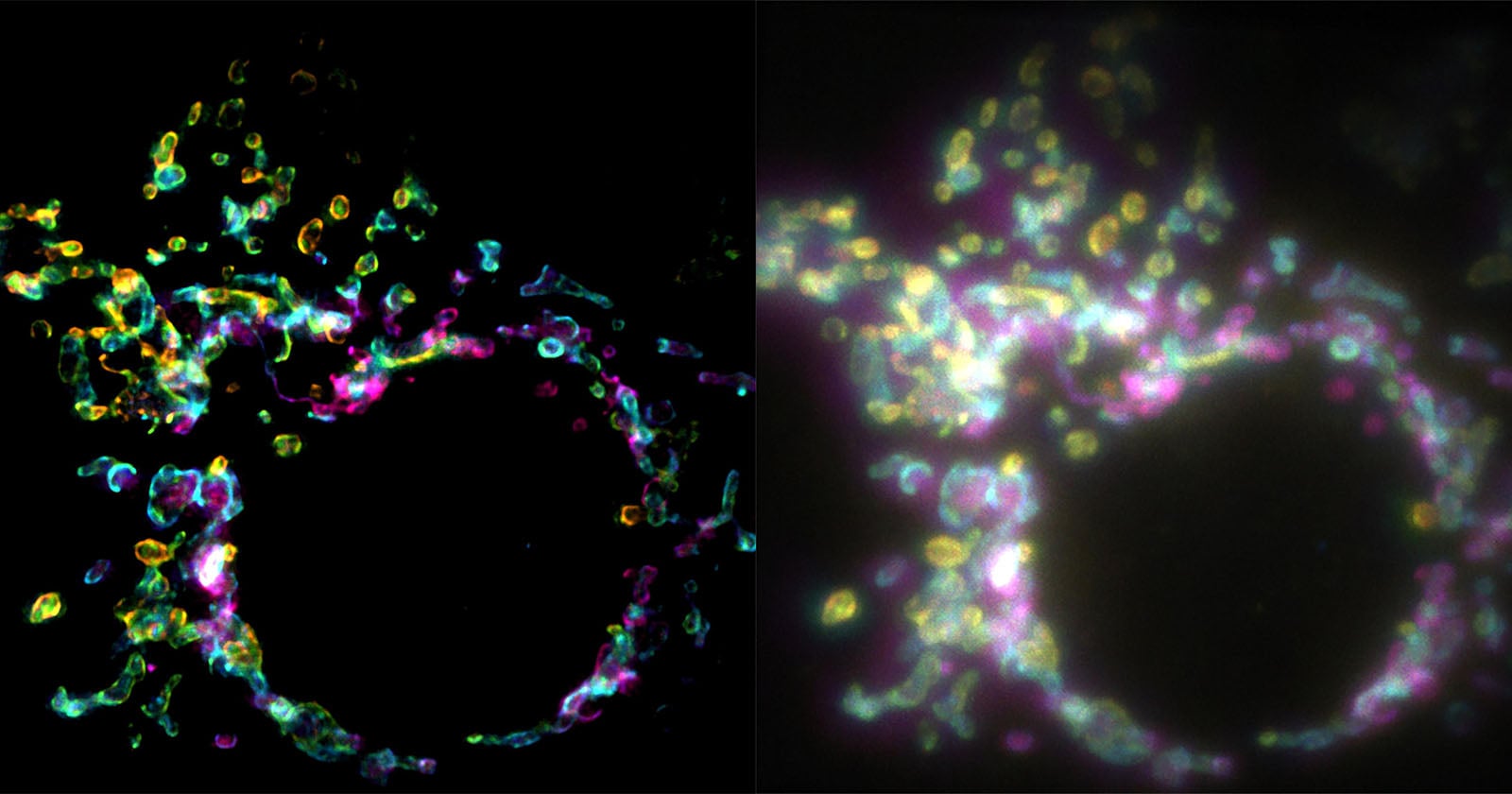
SPIFFI upends this paradigm by shifting the computational burden from temporal data accumulation to spatial polarization analysis. By splitting fluorescent light into four distinct, polarization-sensitive channels simultaneously, the system can reconstruct hidden structural details from just one frame. The technique effectively doubles raw image resolution in a single exposure—resolving structures down to roughly 160 to 170 nanometers—and can be paired with computational post-processing to push thresholds down to an astonishing 80 nanometers. Crucially, the hardware can also be retrofitted onto many existing fluorescence microscopes, promising widespread accessibility across research laboratories worldwide.
Chronology of Discovery
The journey toward SPIFFI began with a fundamental limitation recognized by researchers studying nanoscale biology: cellular processes happen far too quickly for traditional super-resolution techniques to record accurately.
- The Historical Bottleneck: For decades, conventional light microscopy was bound by the diffraction limit of light, restricting resolved details to roughly 200–300 nanometers. While the advent of super-resolution techniques (such as PALM, STORM, and STED) shattered this barrier—earning a Nobel Prize in Chemistry in 2014—they came with a heavy trade-off. They required the collection of extensive time-series datasets. For a living cell undergoing division, migration, or internal trafficking, the subject would shift, morph, or degrade during the prolonged exposure window, resulting in smeared or unusable imagery.
- Conception at EPFL: Recognizing that temporal information was inadequate for live-cell super-resolution, the LBEN team at EPFL—led by Aleksandra Radenovic and spearheaded by PhD student Wei Guo—began exploring alternative physical dimensions of light. Rather than looking at when photons arrived over time, they investigated how the photons were polarized.
- Hardware and Software Integration: Over several years, the team engineered the optical splitter capable of dividing emissions into four polarization-sensitive channels. They developed sophisticated algorithms to compare the angular differences of these channels, effectively turning a single snapshot into a high-density structural map.
- Recent Publication: The culmination of these efforts was formally documented and peer-reviewed, leading to its landmark publication in Nature, instantly generating ripples across the global biophysics community.
Supporting Data and Technical Specifications
To understand the magnitude of the SPIFFI breakthrough, one must examine the quantitative metrics distinguishing it from legacy imaging systems.
- Single-Frame Superiority: Standard fluctuation-based super-resolution imaging requires anywhere from hundreds to tens of thousands of individual frames. SPIFFI achieves super-resolution natively within a single frame. As a result, every single captured frame in a recording is super-resolved, paving the way for true, high-definition super-resolution video of live cellular mechanics.
- Resolution Benchmarks: Out of the box, SPIFFI improves raw optical resolution by a factor of two, successfully resolving intricate structures approximately 160 to 170 nanometers in size. When integrated with existing fluctuation-based post-processing pipelines, the resolution bounds are further refined to approximately 80 nanometers.
- Target Structures: In experimental demonstrations, the research team successfully imaged complex, fast-moving cellular structures, most notably the mitochondrial outer membrane. Mitochondria are notoriously dynamic, constantly fusing, dividing, and shifting positions within the cytoplasm—making them ideal stress-test candidates for real-time super-resolution imaging.
- Hardware Adaptability: Unlike dedicated super-resolution microscopes that require entirely custom-built, multimillion-dollar optical suites, SPIFFI’s optical polarization hardware module can be integrated into pre-existing widefield fluorescence microscopes. This significantly lowers the barrier to entry for biology labs operating on standard institutional budgets.
Official Responses and Researcher Insights
The implications of the SPIFFI technique have been met with immense enthusiasm by its creators and the broader scientific community.
Aleksandra Radenovic, principal investigator at the Laboratory of Nanoscale Biology (LBEN) within EPFL’s School of Engineering, emphasized the transformative nature of the hardware in institutional press statements:
"SPIFFI can capture fast-moving processes within cells, while enabling high-throughput, multi-dimensional imaging beyond the limits of conventional microscopes."
Elaborating on the theoretical shift from older methodologies, first author and LBEN PhD student Wei Guo explained the core frustration of legacy systems that inspired the project:
"Essentially, previous approaches used temporal information to resolve spatial resolution, but this doesn’t work very well on living cells."
Guo further highlighted the revolutionary transition from static imagery to dynamic visual storytelling:
"With previous techniques, taking many images would only result in one super-resolved frame. With SPIFFI, every frame is super-resolved, meaning we can now produce super-resolution videos of live cells. We also seamlessly integrated SPIFFI images with existing fluctuation-based methods for post-processing, achieving resolutions of about 80 nanometers."
Looking forward, the research team is not resting on its laurels. Current developmental efforts are heavily focused on miniaturization. By engineering a more compact, streamlined version of the SPIFFI optical module, the team aims to enhance its day-to-day usability, transforming it from a cutting-edge laboratory prototype into a standardized commercial accessory for biological research hardware.
Implications for Science, Medicine, and Beyond
The introduction of spatial polarization-induced fluorescence fluctuation imaging opens up sweeping new avenues of research across multiple scientific disciplines.
1. Real-Time Virology and Immunology
Understanding how viruses enter cells, hijack cellular machinery, and replicate typically requires watching the infection cycle unfold in real time. Because viral particles and host cellular components operate on nanometer scales and move rapidly, legacy microscopes struggled to capture these interactions clearly. SPIFFI will allow immunologists and virologists to record viral trafficking and immune response mechanisms with unprecedented clarity.
2. Neurobiology and Synaptic Plasticity
Neurons are among the most dynamic and morphologically complex cells in the human body. Processes like axonal transport, neurotransmitter vesicle fusion, and synaptic plasticity occur on sub-second timescales and at the nanoscale. By enabling true super-resolution video, SPIFFI could help neuroscientists map out the physical underpinnings of memory formation, neurodegenerative diseases like Alzheimer’s and Parkinson’s, and cognitive decline.
3. Pharmacology and Drug Discovery
Pharmaceutical research relies heavily on observing how drug candidates interact with cellular receptors, proteins, and organelles. Being able to watch these interactions happen live inside a living cell—rather than inferring mechanics from static post-mortem snapshots or blurry widefield videos—will dramatically accelerate target validation and lead optimization in drug development pipelines.
4. Accessibility and Democratization of Advanced Optics
Perhaps one of SPIFFI’s most understated, yet impactful, implications is its backward compatibility. By ensuring that the hardware can be integrated into standard laboratory fluorescence microscopes, EPFL has avoided the trap of elitist technology. Smaller university laboratories, clinical research centers, and institutions with constrained funding will theoretically be able to upgrade their current microscopes rather than purchasing entirely new imaging suites.
As the EPFL team continues to refine and compact the SPIFFI hardware, the scientific community stands on the precipice of a new visual era. The invisible, hyper-dynamic micro-universe within us is about to come into sharper, clearer focus than ever before.